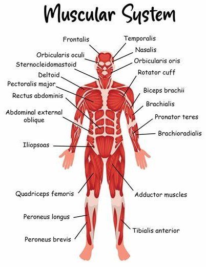
The human body moves because of the complex network of tissues that make up the muscular system. It is made up of three primary muscle types: cardiac, which is only present in the heart and pumps blood throughout the body; skeletal, which attaches to bones and allows voluntary movements; and smooth, which regulates involuntary activities within organs and veins. This system is essential for posture, circulation, and heat production in addition to allowing movement and stability. Gaining an understanding of the muscular system is crucial to understanding how the body functions and reacts to different physical situations and activities.

Table of Contents
MUSCLE
• In muscular systems muscle consists predominantly of canticle cells, produces the moments of various parts of the body by contraction, and occurs in three types:

Skeletal muscular system
- Is considered voluntary and has a striated histologic structure to its component myofibrils.
- Makes up approximately 40% of the total body mass and functions to produce movement of the body, generate body heat, and maintain body posture.
- It consists of two attachments: an insertion, which is normally the more movable and distal attachment, and an origin, which is typically defined by a more stable and proximal attachment.
- Is enclosed by fascia, the epimysium, which is a thin but tough layer of connective tissue sur[1]rounding the entire muscle. Within the muscle, smaller bundles of muscle fibers are surrounded by perimysium. Each individual muscle fiber ls enclosed by endomysium.
Cardiac muscular system
- Is striated muscle fibers found in the wall of the heart, the myocardium.
- Cardiac muscle contractions are autonomous, but the rate can be modulated by the autonomic nervous system (ANS).
- Includes subendocardial specialized myocardial fibers that form the cardiac conducting system.
Smooth muscular system
- Is involuntary and nonstriated and found in the walls of organs and blood vessels.
- In the walls of hollow organs, smooth muscle is arranged in two layers, circular and longitudinal, that allow rhythmic contractions called peristaltic waves in the walls of the gastrointestinal (GI) tract, ureters, uterine tubes, and other organs.
- It is innervated by the autonomic nerve system (ANS), which controls the lumen size of tubular structures.
STRUCTURES ASSOCIATED WITH SKELETAL MUSCLES IN MUSCULAR SYSTEMS

Tendons
- Are fibrous bands of dense connective tissue that conn act muscl&1 ID ban&1 or cartilage.
- Are supplied by sensory fibers extending from muscle nerves.
Ligaments
- Are fibrous bands that connect bones ID bones or cartilage (the term is also used for folds of peritoneum serving to support visceral structures).
Raphe
- is the seam where symmetrical structures, such the scrotal, pharyngeal, and mandibular raphes, unite by a fibrous or tendinous band.
Aponeuroses
- Are flat fibrous tendons of attachment that serve as the means of origin or insertion of a muscle.
Retinaculum
- Is a fibrous thickening of the deep fascia that stabilizes tendons and neurovascular structures as they cross a joint in the distal limbs.
Bursae
- Are fluid-filled flattened sacs of synovial membrane that facilitate movement by minimizing friction between a bony joint and the surrounding soft ti8Sue, such as skin, muscles, ligaments
Synovial tendon sheaths
- Are synovial fluid-filled tabular sacs around muscle tendons that facilitate movement by reducing friction as tendons pass distally into the limbs.
Fascia
- is a fibrous layer that covers the body beneath the skin, penetrates the muscles, and may prevent pus and extravasated fluids like blood and urine from spreading.
1.Superficial fascia
- Is a fatty connective tissue between the dermis and the deep muscular fascia and is considered the hypodermis with fat, cutaneous vessels, nerves, lymphatics, and glands. In a few locations, there may be a membranous deep layer al superficial fascia (abdominal wall).
2.Deep fascia
- is a layer of fibrous tissue that covers the muscles and acts as a stocking or elastic sheath to support them.
- Provides origins or insertions for muscles, forms fibrous sheaths or retinacula for tendons, and forms potential pathways for spread of infection or extravasation of fluids.

